|
Hippocampus
As mentioned many times in the note on Memory and Learning, the key player of memory formation in the brain is Hippocampus. It can never be overemphsized to understand about the details of Hippocampus as much as possible. The more you know about anatomy and functionality of hippocampus, the better you would understand the process of memory formation and recall.
In this section, I would talk about a little bit of details of Hippocampus, but not too details as in academic paper. What I am trying to do is just to give you some keywords and procedure to help you to read documents (e.g, textbook or academic paper).
The first things that I want to suggest you to get familiar with is the basic anatomy of Hippocampus as illustrated below. First you should be able to image the shape and location of Hippocampus in your brain without looking into any reference (e.g, textbook). Then you should be able to identify the location of hippocampus from various cross section images of the brain. And then you should be able to draw (or imagine) cross section of the hippocampus and identify major part of the substructure
of the hippocampus. In short, It would be good enough (at least as an hobbiest) if you can imagine the illustration shown below without looking at this note when you read other document or watching a lecture.
NOTE : as I always says, don't try to memorize it... just read this note or textbook or watch lectures as many as possible and check if you can visualize in your mind without openning the textbook. If you don't get the image in your mind, then look into this note or other textbook.
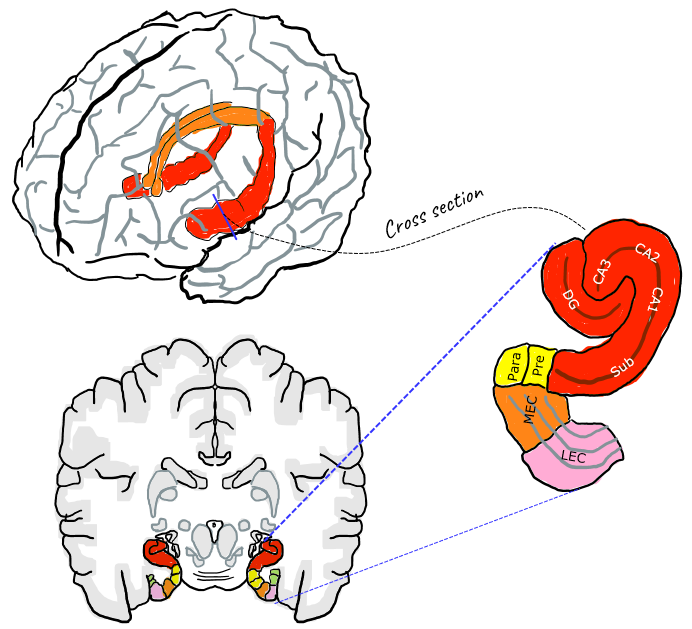
DG : Dentate Gyrus
Sub : Subiculum
Pre : Presuiculum
Para : Parahippocampal gyrus
MEC: medial entorhinal cortex
LEC: lateral entorhinal cortex
Hippocampus Proper = CA1 + CA2 + CA3
Entorinal Cortex = MEC + LEC
Following is the overal circuitary connecting various part of substructure of Hippocampus and cortex of the brain. I know it look confusing. Don't try to memorize it. Just to try to follow one or two specific circuits when you have chance (e.g, reading other textbook or sitting in a lecture).
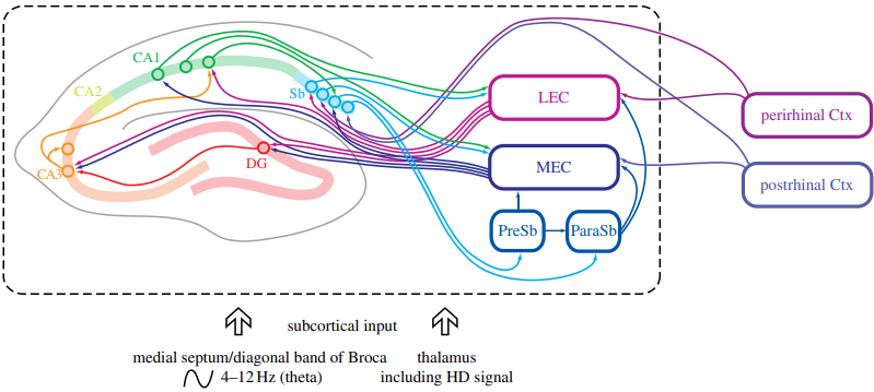
Image Source : Space in the brain: how the hippocampal formation supports spatial cognition
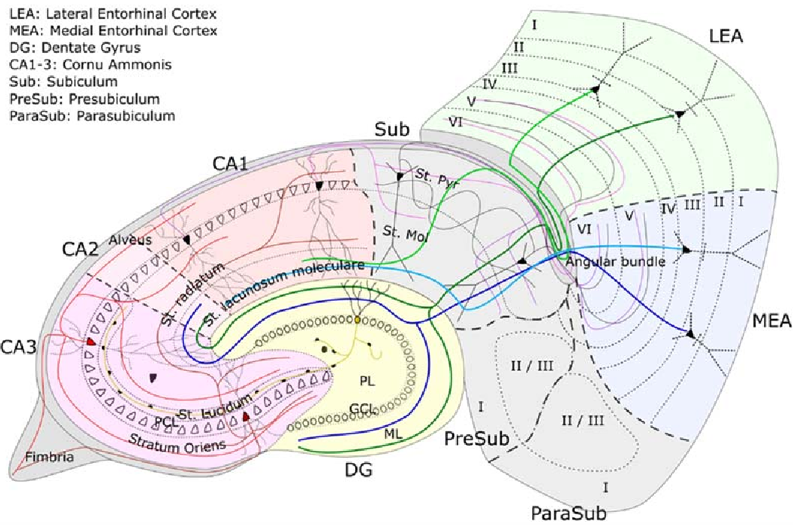
Image Source : Marr’s theory of the hippocampus as a simple memory
Followings are the summary of functionalities of each subregions in Hippocampus : (Mostly based on Exploring the Function of the Hippocampus by Anatomy )
Dentate Gyrus :Dendate Gyrus plays various functionas as below
- is involved in the mnemonic (memory) processing of spatial information, the formation of episodic and spatial memories, and the spontaneous exploration of novel environments
- is the input region of the hippocampus, and serves as a pre-processing unit
- some information from the entorhinal cortex directly enters the dentate gyrus, is processed by dentate gyrus, and then the processed information is delivered to the CA3 region of the hippocampus proper
- perform pattern separation, which is separating relatively similar input patterns into distinct/unique output patterns
- is required for Working Memory & Spatial Navigation
- Undergoes LTP and Neurogenesis
- Required for Working Memory & Spatial Navigation
CA1 : CA1 is the first region of Hippocampus in terms of anatomy and plays various functions as follows.
- Recieves inputs from CA3 or CA2 and output the information to entorhinal cortex
- (more specifically) CA1 recieves temporal information from CA2 and spatial information from CA3.
- one of the functions of the CA1 region may be to produce combined spatial-temporal code that allows an animal to distinguish the memory of 2 events from each other, even if they occur at the same place but in different times.
- Performs the retrieval of episodic autobiographical memory
CA2 : CA2 is located between CA1 and CA3 anatomically and plays various functions as follows.
- Neurons in this region carries less spatial information than in CA1 or CA3.
- receives input from layer 2 of the entorhinal cortex through the perforant pathway
- encodes time information more strongly than spatial information and then send the temporal information to CA1
- has a function pertaining to social behavior and may be involved in social memory processing (e.g, differentiating between new and familiar individual around you)
CA3 : CA3 is located farthest away from CA1 and sitting right next to DG(Dentate Gyrus).
- receives input from both the mossy fibers of the granule cells in the dentate gyrus, and from cells in the entorhinal cortex through the perforant pathway.
- considered to be the “pacemaker” of the hippocampus, and thereby has a big role in learning
- operated as an autoassociation memory to store episodic memories including object and place memories
- dentate granule cells operated as a preprocessing stage for the CA3 region by performing pattern separation so that the mossy fibers could act to set up different representations for each memory to be stored in the CA3 cells.
- the mossy fiber system to CA3 connections are involved in learning but not in recall
- involved in the acquisition of context-dependent fear extinction
Subiculum :Subiculum is between CA1 and Parahipocampal gyrus and plays roles as follow.
- Functions as main output port of Hippocampus
- has functions pertaining to memory, spatial navigation, mnemonic or symbol processing, and regulating the body’s response to stress through the inhibition of the HPA axis.
- Responds to an influx of information as well (e.g,to only familiar objects or a landmarks, and to concurrent location and speed)
Entorhinal Cortex :Entorhinal Cortex (MEC + LEC) located in the medial temporal lobe and is next to the hippocampus
- Functions as both the major input and the major output hub of information to and from the hippocampus
- Interface between the neocortex and the hippocampus
- (with the hippocampus) Works for formation on declarative/explicit (autobiographical, episodic, semantic) memory storage, and spatial memories in particular.
- Collaborates with hippocampus on memory formation, memory consolidation, and memory optimization in sleep.
- (with the hippocampus) plays key roles for our sense of direction and spatial navigation ability
Looking in a little bit of broader scope regarding the overall neural path for memory encoding and decoding. It is nicely summarized in illustrated as below.
During memory encoding, visual and other sensory information are processed by the dorsal and ventral streams, integrated in the PHC and PRC, and sent to the EC. The EC then transmits the information through the hippocampal trisynaptic circuit (DG, CA3, and CA1), where it is further processed and consolidated. The subiculum sends the processed information back to the EC and other cortical areas for long-term storage.
During memory retrieval or decoding, the EC retrieves stored information and sends it back to the hippocampus. The hippocampus reconstructs the memory representation through pattern completion, and the subiculum sends the retrieved information to cortical areas for conscious awareness and further processing.
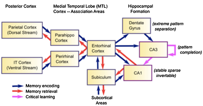
Image Source : The extended trajectory of hippocampal development: Implications for early memory development and disorder
Dorsal and Ventral Streams: Visual information enters the brain through the retina and is processed in the primary visual cortex (V1). From there, it is sent to two parallel pathways: the dorsal stream, responsible for spatial processing and motion (the "where" pathway), and the ventral stream, responsible for object recognition and form (the "what" pathway). Both streams contribute to memory formation by processing and transmitting different aspects of visual
information.
Parahippocampal Cortex (PHC): The PHC receives input from the dorsal and ventral streams and plays a crucial role in processing contextual and spatial information. It is involved in encoding and retrieving contextual associations in memory.
Perirhinal Cortex (PRC): The PRC receives input primarily from the ventral stream and is involved in object recognition and associative memory. It contributes to the encoding and retrieval of object-related information and helps integrate this information with other sensory modalities.
Entorhinal Cortex (EC): The EC receives information from the PHC, PRC, and other cortical areas. It plays a critical role in connecting the neocortex to the hippocampus and serves as a gateway for information flow. The EC contains grid cells, which help form spatial representations and are essential for spatial memory.
Dentate Gyrus (DG): The DG receives input from the EC and is the first stage of the hippocampal trisynaptic circuit. It plays a role in pattern separation, which helps create distinct memory representations and reduces interference between similar memories.
CA3: The DG sends information to the CA3 region, which is involved in associative memory processes and pattern completion. CA3 has extensive recurrent connections that enable the retrieval of complete memory representations even with partial input.
CA1: Information from CA3 is sent to the CA1 region, which is essential for the encoding and retrieval of episodic memories. CA1 integrates information from CA3 and direct input from the EC, sending the processed information back to the EC and other cortical areas.
Subiculum: The subiculum receives input from CA1 and serves as the main output structure of the hippocampus, sending information back to the EC, PHC, PRC, and other cortical areas. The subiculum is involved in spatial memory and processing spatial context.
CA2 : The CA2 region, located between CA1 and CA3, has unique properties and is involved in social memory and the encoding of novel experiences.
Experts keep saying (it is well known) that Hippocampus plays the most crucial roles in the memory process. With all the technical adances and creative ideas(sometimes looking like pure science fiction), you may ask 'Can we make any silicon chipset or computer program that can emulate/simulate Hippocampus ?'.
Motivation
Why we want to duplicate Hippocampus instead of trying to duplicate other area of brain cortex ?
The hippocampus is relatively well-understood in terms of its structure and function, particularly in relation to memory formation, compared to the more complex and less localized functions of the broader cerebral cortex. It is one of the most studied areas of the brain in neuroscience due to its clear involvement in the formation of new memories and its relatively distinct anatomy. The hippocampus' processes are somewhat more defined and discrete, making it a more approachable target for replication
and prosthetic development. In contrast, memories are believed to be stored diffusely across the cerebral cortex, which makes the replication of those cortical areas a more complex and less defined task.
Here goes a list of possible motivation behind this idea
- Restoring memory functions lost due to diseases or injuries.
- Understanding and modeling brain functions to advance neuroscientific knowledge.
- Developing prosthetic devices that can bypass damaged neural areas.
- Creating systems to simulate and predict brain behavior for research purposes.
- Contributing to the development of artificial intelligence by mimicking biological processes.
- The hippocampus is the most ordered and structured part of the brain, and one of the most studied. Importantly, it is also relatively easy to test its function.
Case Stories
- Duplicating CA3 with VLSI : This is based on VLSI Implementation of a Nonlinear Neuronal Model: A “Neural Prosthesis” to Restore Hippocampal Trisynaptic Dynamics
- The research aims to substitute the biological function of the CA3 region in the hippocampus of a rat with a VLSI (Very Large Scale Integration) chip to restore memory function potentially lost due to diseases like Alzheimer's.
- The project involves surgically removing CA3 function and using a multi-site electrode array to transmit signals from the dentate gyrus (DG) to the VLSI chip and then to the CA1 region, effectively bypassing the damaged CA3.
- The results demonstrated that the VLSI chip could replicate the biological propagation of signals in the hippocampus, showing promise for this type of neural prosthesis in memory restoration.
- World's first brain prosthesis : This is based on World's first brain prosthesis revealed
- Model, build, interface
- Creating a mathematical model of the hippocampus's function under various conditions. : Studying rat hippocampus slices, observing their response to electrical stimulation and Developing a model from these observations.
- Programming this model onto a silicon chip.
- Interfacing the chip with the brain via electrode arrays : Placing the chip on the skull, connected to electrodes on either side of the damaged area.
- Testing
- Initial tests will be on rat brain slices in cerebrospinal fluid.
- If successful with rats, live rat and monkey tests are planned, focusing on memory tasks.
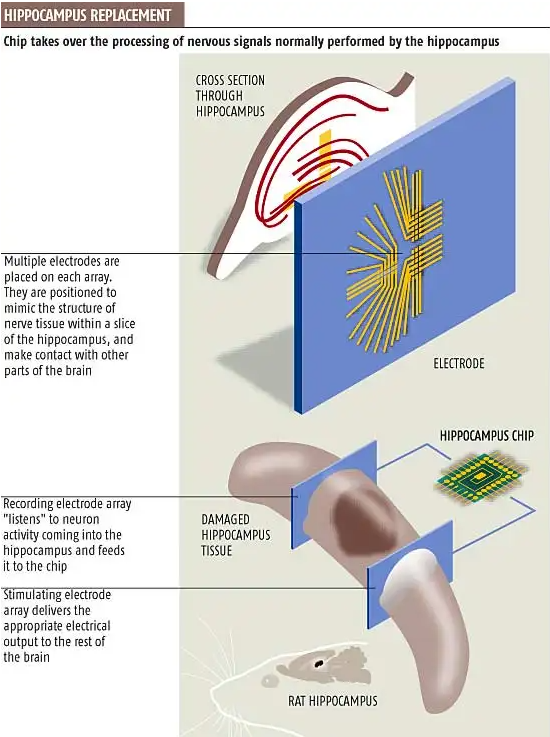
Image Source : World's first brain prosthesis revealed
YouTube
- Milestone Research / Observation
- Process of Memory/Learning
- Place / Navigation
- Reliability of Memory
- Inheritance of Memory
- Lectures
- Efficient Learning
- Pioneering Researches
- Further Watching
- World Science Festival
Reference
- Well-known persons
- Memory Process
- Type of Memory
- Reliability Of Memory
- Mechanism of False Memory / Confabulation / Lie
- Inheritance of Memory
- Spatial learning / Navigation
- Place / Grid Cells involving in Non spatial cognitive processing
- Schema
- Cognitive Load
- Dual process theory
- Knowledge
- Anatomy
- Pioneering Researches
|
|


